


3D printing has a lot of use cases for example it is used in medicine and in schools. So far skeletons for anatomy classes were produced with conventions techniques but that’s changing in the last time.
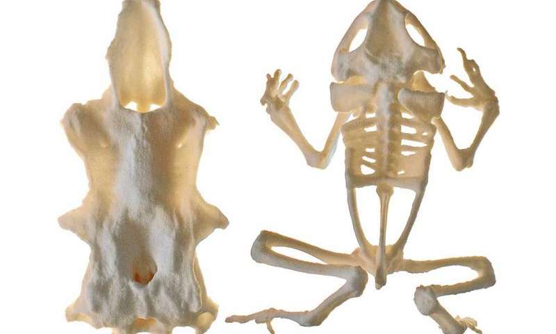
3D printed models of skeletons of the human body are already used in the last time. These models are used by students to learn anatomy. They are a cheaper alternative to conventional produced skeletons and to real skeletons. The new project is lead be Dr. Daniel Thomas from the Massey University in New Zealand. Target of the project is to scan the skeleton of the cane toad (Rhinella marina) and spiny dogfish (Squalus acanthias). For the scanning they used a NextEngine HD desktop scanner and a David SLS-2 structured light scanner. To print the scanned objects they used a 3D printer that’s suited up for the SLS technique.
Anatomy teaches us about the ecology and evolution of an animal and can give us crucial information for developing conservation strategies. It’s not always possible for learners to study original anatomy specimens though, which is where high-quality 3D printed models come in.
The project will be continued to scan as much animals as possible. Dr Daniel Thomas has also announced that printing bones larger than the maximal build size of the 3D printer is no problem. To make it possible, the bones are spitted up and connected after 3D printing.
Subscribe to our Newsletter
3DPResso is a weekly newsletter that links to the most exciting global stories from the 3D printing and additive manufacturing industry.