


Scientists at the Wyss Institute of Harvard University have used 3D bioprinting to create tubular kidney architecture that mimics the function of a human kidney.
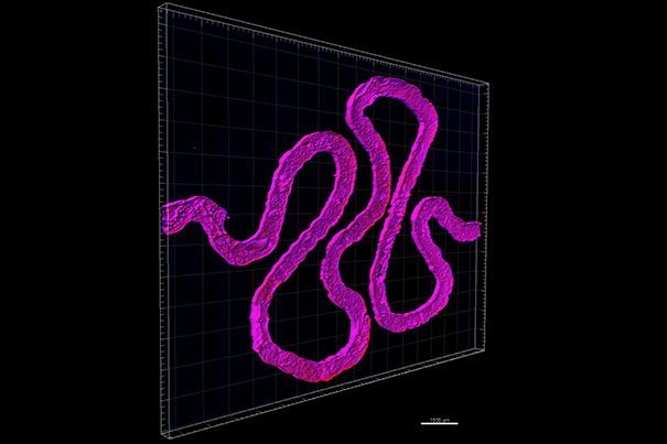
The current research builds upon the successful results to 3D print thick vascularised tissue, that was viable for more than one month in vitro. In collaboration with Roche scientist Annie Moisan, Jennifer Lewis and her team created a functional 3D renal structure with living human epithelial cells, that line the kidney tubules’ surfaces. It mimics a hollow tube, called proximal tubule, that is an essential part of each of the over one million nephrons in every human kidney. Proximal tubules are responsible for reabsorbing nutrients that are transported back into the bloodstream.
“The current work further expands our bioprinting platform to create functional human tissue architectures with both technological and clinical relevance,” Lewis explains.
Co-first authors of the study Kimberly Homan and David Kolesky emphasised that the model is more than just mimicking a proximal tubule. It is “a credible in vitro model that functions like living kidney tissue, representing a significant advance from traditional 2D cell culture.”
The extracellular matrix base layer was created by using a 3D printed silicone gasket as a mould. A fugitive ink was then printed in a convoluted winding tubular shape and encapsulated with another layer of extracellular matrix. The fugitive ink is then liquefied and removed from the final architecture.
“The use of functional tissue-like models during pre-clinical studies will provide unprecedented insights into human-relevant drug response prior to clinical development,” added Moisan, Laboratory Head in Mechanistic Safety at Roche and author of this study.
Eventually these models could be used for implants or organ-assitive devices.
Subscribe to our Newsletter
3DPResso is a weekly newsletter that links to the most exciting global stories from the 3D printing and additive manufacturing industry.