Algerian scientists have conducted a study on the production of anatomical bone models using 3D printing. The aim was to create cost-effective and realistic illustrative objects for teaching anatomy in veterinary faculties.
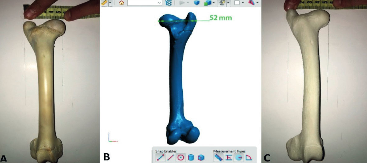
The researchers from the School of Veterinary Medicine in Algiers chose the right femur of a sheep as the starting point. After preparing the bone, 3D digitization was carried out using a surface scanner. The scan data was converted into a virtual 3D model.
An SLS Spro60 HD 3D printer from 3D Systems was then used to create the femur model from polyamide PA12 using selective laser sintering. “The precision of the 3D printer was calibrated to an accuracy of 0.069 mm,” says a researcher, explaining the process.
In the next step, the printed bone was reworked to achieve the most realistic surface structure possible. The analysis showed that all anatomical details were accurately reproduced, apart from minor deviations in the depth of the sewing hole.
A comparison of the dimensions between the original bone, virtual model and printed model showed only minimal differences of a maximum of 1 mm. “In view of this small error of 1 mm, 3D-printed models can certainly serve as a reliable alternative to real bones in anatomy lessons,” the scientists summarize.
The paper entitled “Comparative study between bone obtained by 3D printing and its original model” can be accessed here.
Subscribe to our Newsletter
3DPResso is a weekly newsletter that links to the most exciting global stories from the 3D printing and additive manufacturing industry.


