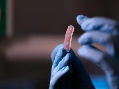
Researchers at Harvard’s John A. Paulson School of Engineering and Applied Sciences (SEAS) have developed a method to create a beating heart tissue using 3D printing technology. By using a novel hydrogel ink enriched with gelatin fibers, the scientists were able to create a functioning heart ventricle model that behaves similarly to a human heart.
3D printing has opened up new possibilities for bioengineers* to create heart tissues and structures over the past decade. Researchers* have now found a way to print heart muscle cells so that they align and beat like a human ventricle.
“People have been trying to replicate organ structures and functions to test drug safety and efficacy as a way of predicting what might happen in the clinical setting,” says Suji Choi, research associate at SEAS and first author on the paper.
But until now, 3D printing techniques alone have not been able to achieve physiologically relevant alignment of cardiomyocytes, the cells responsible for coordinated transmission of electrical signals to contract the heart muscle.
“We started this project to address some of the inadequacies in 3D printing of biological tissues”, states Kevin “Kit” Parker, Tarr Family Professor of Bioengineering and Applied Physics, head of the Disease Biophysics Group at SEAS, senior author.
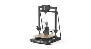
“FIG ink is capable of flowing through the printing nozzle but, once the structure is printed, it maintains its 3D shape,” says Choi. “Because of those properties, I found it’s possible to print a ventricle-like structure and other complex 3D shapes without using extra support materials or scaffolds.”
To make the FIG ink, Choi used a rotary-jet spinning process developed by Parker’s lab that can spin microfiber materials similar to cotton candy. Postdoctoral researcher Luke MacQueen, a co-author of the paper, came up with the idea that the fibers made using the rotary jet process could be added to an ink and 3D printed.
“When Luke developed this concept, the vision was to broaden the range of spatial scales that could be printed with 3D printers by dropping the bottom out of the lower limits, taking it down to the nanometer scale,” Parker says. “The advantage of producing the fibers with rotary jet spinning rather than electrospinning” – a more conventional method for generating ultrathin fibers – “is that we can use proteins that would otherwise be degraded by the electrical fields in electrospinning.”
Using the rotary nozzle to spin gelatin fibers, Choi created a sheet of material that looks similar to cotton. Next, she used sound waves to break this sheet into fibers 80 to 100 micrometers long and 5 to 10 micrometers in diameter. She then dispersed these fibers into a hydrogel ink.
The biggest challenge was finding the optimal ratio of fibers to hydrogel in the ink to ensure fiber alignment and the integrity of the 3D-printed structure.
When Choi printed 2D and 3D structures with the FIG ink, the cardiomyocytes in the ink aligned with the direction of the fibers. Thus, by controlling the direction of printing, Choi could control how the cardiomyocytes would align.
This invention offers an exciting prospect for creating in vitro platforms for discovering new therapeutics for heart disease and evaluating the effectiveness of treatments in individual patients.
Additional authors include Keel Yong Lee, Sean L. Kim, Huibin Chang, John F. Zimmerman, Qianru Jin, Michael M. Peters, Herdeline Ann M. Ardoña, Xujie Liu, Ann-Caroline Heiler, Rudy Gabardi, Collin Richardson, William T. Pu, and Andreas Bausch.
Subscribe to our Newsletter
3DPResso is a weekly newsletter that links to the most exciting global stories from the 3D printing and additive manufacturing industry.


